
Have you ever wondered what lies beneath the slimy skin of a frog? With a little bit of knowledge and a steady hand, you can dissect a frog and unravel its fascinating inner organs. Frogs are commonly dissected in biology classes to understand their anatomy and how different organs work together to help them survive in their natural habitat.
By dissecting a frog, you can explore its skeletal structure, muscular system, circulatory system, digestive system, and even its reproductive organs. It’s a hands-on experience that allows you to visualize and understand the intricate workings of a living creature. Whether you are a student or simply curious, dissecting a frog can be an enlightening and educational experience.
Not only can dissecting a frog help you understand its anatomy, but it can also teach you valuable skills such as observation, patience, and hand-eye coordination. It’s an opportunity to apply what you have learned in the classroom to a practical setting. By using tools like scalpels, forceps, and scissors, you can carefully examine each organ and understand its purpose within the frog’s body.
Getting Started: Preparing for the Dissection
- Creating a Clean Workspace: A clean and organized workspace is essential for conducting a successful dissection. Clear away any clutter and ensure that all surfaces are clean and disinfected. Make sure to have proper ventilation in the room to avoid any unpleasant odors.
- Obtaining the Frog: A frog can be obtained from a scientific supply company or a local biology lab. Ensure that the frog is properly preserved and stored before beginning the dissection.
- Thawing the Frog: If the frog is frozen, it needs to be thawed prior to the dissection. This can be done by placing the frog in a refrigerator overnight or by following the instructions provided with the preserved frog.
- Positioning the Frog: Once the frog is thawed, it should be positioned properly in the dissecting pan. The ventral side of the frog, which is the side with the belly, should face up for easy access to the internal organs.
- Ready, Set, Dissect: With all the necessary preparations done, it is time to begin the dissection. Follow the step-by-step instructions outlined in a dissection manual or guide, ensuring precision and accuracy throughout the process.
Obtaining a Frog

Purchasing a Preserved Frog
If you choose to purchase a preserved frog, there are several options available. You can find preserved frogs online or at scientific supply stores. Make sure to choose a high-quality specimen that has been properly preserved. It is also important to consider the size of the frog, as larger frogs are generally easier to dissect and examine.
Capturing a Frog from the Wild
If it is legal and ethical to capture a frog from the wild in your area, there are a few methods that can be used. One method involves setting up traps near bodies of water where frogs are commonly found. These traps can be baited with food, such as insects or small pieces of meat, to attract the frogs. Once a frog enters the trap, it can be carefully captured and transferred to a suitable container for transport.
It’s essential to handle the frog with care and respect throughout the process. Always remember to consider the well-being and conservation of these creatures. Once you have obtained a frog, you can proceed with the necessary preparations for the dissection.
Gathering Necessary Tools and Equipment for a Frog Dissection
1. Dissection Kit: A good quality dissection kit is essential. It typically includes a scalpel, forceps, scissors, and probes. These tools allow you to make precise incisions, handle tissues, and explore various body parts of the frog.
3. Safety Glasses: Safety glasses or goggles should be worn at all times during the dissection to protect your eyes from any potential splashes or spills of fluids.
4. Dissection Tray: A flat and sturdy dissection tray is necessary to securely hold the frog during the dissection. It should be made of a material that is easy to clean and disinfect.
5. Preserved Frog: A preserved frog is needed for the dissection. It is typically obtained from a scientific supplier and has been specially prepared with preservatives to maintain its anatomical structures.
6. Plastic Bags or Ziplock Bags: These bags are useful for storing any dissected frog parts or materials that are not needed immediately.
7. Dissection Pins: Dissection pins are used to secure the frog onto the dissection tray and hold its body parts in place during the dissection process.
8. Paper Towels: Paper towels are handy for wiping off excess fluids, cleaning the tools, and keeping the work area tidy.
9. Disposable Containers: Disposable containers are useful for holding any fluids or specimens that are extracted during the dissection.
Having these tools and equipment readily available will ensure that you can confidently and efficiently carry out a frog dissection. Remember to handle all tools and materials with care and follow proper safety protocols throughout the process.
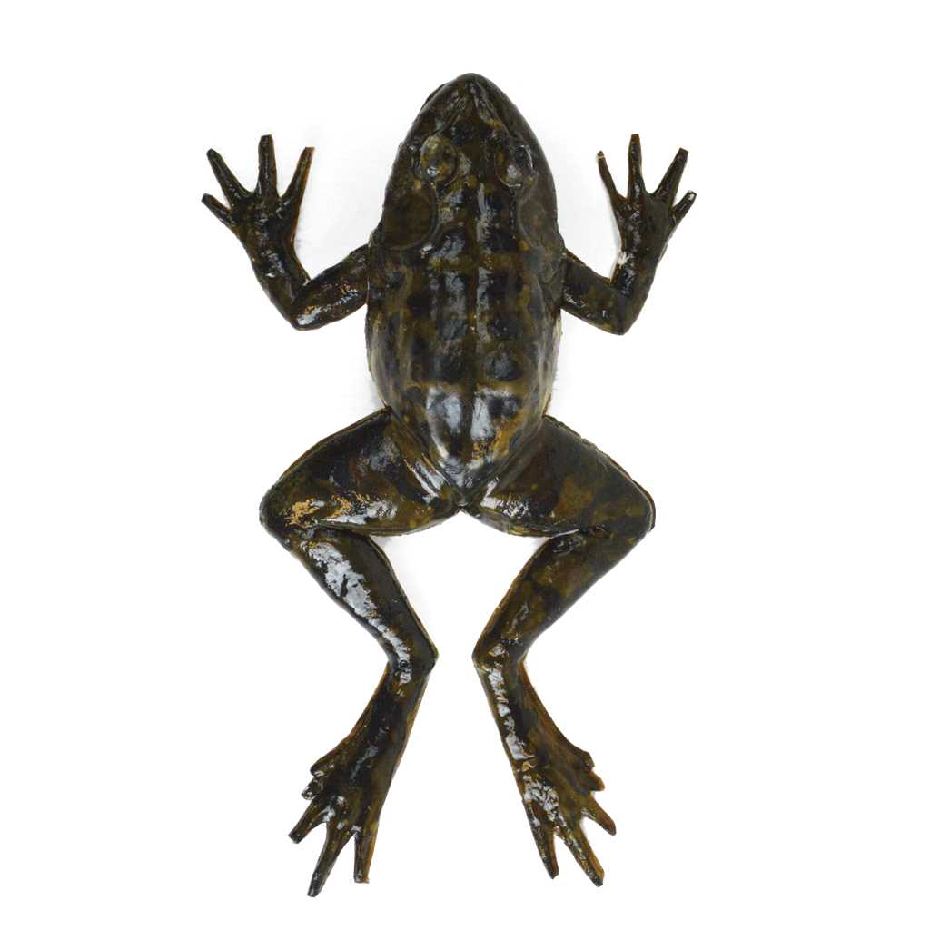
Exploring the Surface: External Anatomy of a Frog
Start by observing the frog’s overall body structure. Notice its streamlined shape, designed for efficient movement both in water and on land. Take note of its four limbs, which allow it to swim, hop, and leap with agility. These limbs are equipped with digits that aid in gripping and climbing. Additionally, you may notice webbed feet adapted for swimming and jumping.
The skin of a frog is another interesting aspect to examine. Not only does it help retain moisture in terrestrial environments, but it also facilitates respiration as a frog can absorb oxygen through its skin. This unique ability allows a frog to breathe in both water and air, making it a truly remarkable organism.
As you further explore the external anatomy, you will come across various surface features that serve specific functions. For instance, the eyes of a frog are positioned on top of the head, providing a wide field of vision and enabling it to detect prey and predators from different angles. The tympanic membrane of a frog, located just behind the eye, acts as an eardrum and allows it to hear environmental sounds.
Additionally, a frog’s mouth consists of a hinged jaw with a sticky tongue, which it can extend to catch insects and other small prey. Its nostrils are positioned on the snout and enable it to breathe while partially submerged in water.
By closely examining the external anatomy of a frog, you can begin to appreciate the intricate adaptations that allow it to thrive in its environment. From its streamlined body shape to its versatile limbs and unique skin structure, every aspect serves a purpose in the frog’s survival, illustrating the wonders of nature and the incredible diversity of life on Earth.
Examining the Body Features

Skin: The skin of a frog is smooth and moist, providing protection against dehydration and allowing for easy movement in water. It is covered in mucus, which helps to keep the skin moist and is also important in respiration.
Limbs: Frogs have four limbs, which are adapted for different purposes. The forelimbs are shorter and more muscular, allowing the frog to leap and land effectively. The hindlimbs are longer and built for propulsion, enabling the frog to swim and jump long distances.
Eyes: The eyes of a frog are protruding and positioned on the sides of the head, providing a wide field of vision. They are adapted for both vision in air and underwater, with a protective membrane that allows for clear underwater vision.
Mouth: The mouth of a frog is wide and equipped with a long, sticky tongue. This enables the frog to accurately capture and consume its prey, which mostly consists of insects and small vertebrates.
Nostrils: Frogs have two nostrils located on the upper surface of the head. These nostrils are used for breathing and olfaction.
Identifying the External Organs
Skin
The skin of a frog is smooth and moist, providing protection against dehydration. It is also responsible for gas exchange, allowing the frog to breathe through its skin.
Eyes
The eyes of a frog are located on the front of its head, providing excellent binocular vision. Frogs have a nictitating membrane, a transparent inner eyelid that protects their eyes while underwater.
Ears
Frogs have two external ear openings called tympanic membranes or eardrums. These membranes allow them to detect sounds and vibrations in the environment.
Nostrils
Mouth
The mouth of a frog contains several important structures, including the maxillary teeth, vomerine teeth, and a sticky tongue. Frogs use their mouths to catch and swallow prey.
Limbs
A frog has four limbs, each with five digits. The front limbs are adapted for grasping and landing, while the hind limbs are powerful and enable the frog to jump and swim.
By familiarizing yourself with these external organs, you can better understand the frog’s physical adaptations and how they contribute to its survival.
Dissecting a frog can be a can incredibly educational and fascinating hands-on experience. By carefully examining the external organs, you can gain a greater appreciation for the complexity of the frog’s anatomy and its adaptation to its environment.
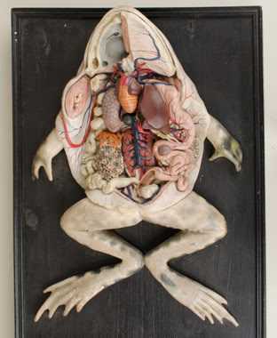
Internal Anatomy: Investigating the Organs
Once the frog has been obtained and the necessary tools and equipment have been gathered, it is time to move on to the internal anatomy of the frog. This is where the true exploration and learning begins.
Respiratory System
Circulatory System

The circulatory system of the frog consists of the heart, blood vessels, and blood. By dissecting the frog, you can locate and examine the heart, observe how blood vessels branch out from it, and even see the different chambers of the heart.
Digestive System
The digestive system of the frog includes the mouth, esophagus, stomach, small intestine, large intestine, and anus. By dissecting the frog, you can follow the path of food as it moves through the digestive system and observe the different organs involved in the process.
Remember, this is an opportunity to learn and explore, so take your time and observe the different organs and structures carefully. Make sure to ask questions and seek guidance if needed. With patience and curiosity, you can uncover the secrets of the frog’s internal anatomy.
Opening the Frog: Initiating the Dissection
Once you have gathered all the necessary tools and equipment, it is time to start the dissection process. Opening the frog is the first step in exploring its internal anatomy. This is a crucial step as it allows you to examine the organs and systems within the frog’s body. The key is to handle the frog with care and precision to ensure a successful dissection.
Step 1: Positioning the Frog
To start, place the frog on a dissecting tray with its ventral side facing up. Make sure the frog is securely positioned so that it doesn’t move during the dissection. This will give you a stable surface to work on and ensure proper visualization of the internal structures.
Step 2: Obtaining the Necessary Tools
Before proceeding, double-check that you have all the tools required for the dissection. These typically include scissors, forceps, a scalpel or a razor blade, and a probe. Having these tools readily available will make the dissection process smoother and more efficient.
Step 3: Making the Incisions
Using the scissors or scalpel, carefully make a midline incision along the ventral side of the frog. Start from the cloaca and extend it towards the throat. Take your time and work slowly to avoid damaging the internal organs. As you make the incision, be mindful of any structures you encounter.
Step 4: Peeling Back the Skin
Once the incision is made, use the forceps to gently lift and peel back the flaps of skin on both sides. Take care not to tear or damage the underlying organs. If needed, use the probe to separate and expose the organs for better visibility.
Step 5: Exploring the Cavity
Step 6: Identifying and Removing the Glands
Pay close attention to any glands you come across during the dissection. This includes the thymus gland and the fat bodies. Take note of their location and appearance. If desired, carefully remove these glands for further examination.
Step 7: Documenting and Observing
Throughout the dissection process, it is essential to document your observations. Take clear and detailed notes of what you see and any significant findings. Additionally, take photographs if possible to create a visual record of the dissection.
Exploring the Digestive System
How is the Digestive System Organized?
The frog’s digestive system can be divided into several main parts, each with its own unique functions. These parts include the mouth, esophagus, stomach, small intestine, large intestine, and cloaca. Each organ plays a vital role in the digestion and absorption of nutrients.
What Can You Discover?
By dissecting a frog, you can observe the different organs that make up the digestive system and examine how they are connected. You can also examine the shape, size, and texture of each organ to better understand its function.
You will be able to see the mouth, which is equipped with a sticky tongue used to capture prey. The esophagus, a muscular tube, connects the mouth to the stomach, where food is broken down with the help of various enzymes.
In the small intestine, the majority of nutrient absorption takes place. The large intestine then processes waste material before it is eliminated through the cloaca, the common opening for the digestive, reproductive, and urinary systems.
Studying the frog’s digestive system can be beneficial for several reasons. Firstly, it provides insight into the similarities and differences between the digestive systems of different organisms, including humans.
Observing the Circulatory System
When a frog is dissected, one of the important systems to observe is the circulatory system. This system plays a crucial role in the frog’s overall functioning, as it allows for the transportation of oxygen, nutrients, and waste products throughout the body.
To examine the circulatory system, it is necessary to carefully remove the frog’s organs and tissues to gain access to the blood vessels. The cardiovascular system of the frog consists of a heart, arteries, veins, and capillaries, all of which work together to ensure the proper circulation of blood.
1. The Heart: The frog’s heart is located in the chest cavity just below the throat. It is a small, muscular organ that pumps blood throughout the body. The heart consists of three chambers: two atria and one ventricle. The atria receive blood from different parts of the body, while the ventricle pumps blood out to the rest of the body.
3. Veins: Veins are blood vessels that carry deoxygenated blood back to the heart. It is essential to locate and trace the major veins, including the vena cava, which is the largest vein returning blood to the heart. The vena cava collects deoxygenated blood from various parts of the body and empties it into the right atrium of the heart.

I’m Lena Adams—a product of an unconventional upbringing in the African wilderness. My father, a daring explorer of African wildlife, sparked my fascination with reptiles, a passion that intertwined with the tragic loss of my mother during an expedition, leaving an indelible mark on my life. Driven to understand the creatures that captivated my parents, I embarked on my journey, sharing insights about reptiles, frogs, and lizards on my website. Through my explorations and conservation efforts, I honour my family’s legacy while seeking connections—to the creatures, nature, and the mother whose presence I yearn to understand.